
On Bartonella Infection

Disclaimer: This blog post is based on my experiences as a patient. I am on a long-haul journey to find answers and resolution to a chronic condition that I have suffered with for 11 years. I am not medically trained. While I have researched this subject matter carefully, this post does not provide medical advice. If you are suffering from symptoms that you think a chronic infection may be the cause of, then please consult a medical professional.
Introduction
Bartonella infection (Bartonellosis) is an infection that biting insects, such as fleas, ticks, or lice, transmit to humans. Contaminated bites and scratches, such as cat scratches can also transmit Bartonella infection. ‘Bartonella infection causes severe clinical syndromes in humans. The three pathogens responsible for the majority of Bartonella infections are B henselae, B quintana, and B bacilliformis.’ [1]
There are three subdivisions of the Bartonella infection: 1) Carrion’s disease, which Bartonella bacilliformis causes. 2) Cat scratch disease (CSD) (Bartonella henselae); and 3) Trench fever (Bartonella quintana). Culture, serology, or histopathology establish the laboratory diagnosis of Bartonella infection.

Somebody on X (formerly Twitter) has reached out to tell me that he thinks that there is a 90% chance that I have a Bartonella infection. It was the contact that I had with this person that inspired me to write this blog post. This post examines what a Bartonella infection is and discusses its causation, symptoms, and the main investigations that doctors use to identify it. It also highlights the congruence between the Bartonella henselae form of Bartonella infection (cat scratch disease) and my atypical symptoms, particularly the chronicity of my condition and the undetected aetiology. This post asks: Do I have a Bartonella infection?
Investigations.

The investigations that clinicians order to test for Bartonella infection are:
- serology: Bartonella henselae (cat-scratch disease [CSD])
- culture: B henselae (CSD)
- aspiration or biopsy of lymph nodes (CSD)
- serology: B quintana (trench fever) [2]
I have written a lot about my 11-year diagnostic delay. I speak about a common pattern over these 11 years, with medics ignoring my blood test results. This has been most common when my neutrophils and white cell count have been abnormal. My additional vexation is that medics have not carried out blood tests, which identify rare infectious diseases, like a Bartonella infection.
Doctors have ordered many blood tests to monitor my full blood count. However, they haven’t done any blood tests to look for specific infectious diseases, apart from two Lyme disease tests that I had back in 2013. They have never done a serology for Bartonella henselae. I have neither had an aspiration nor a biopsy of my lymph nodes. After my parotid surgery in 2018, clinicians missed a major opportunity. A maxillofacial surgeon removed some of my parotid tissue, in which they found dense secretions in the ducts, calcified tissue, and reactive lymph nodes (lymphadenopathy). However, they did not culture my lymph node tissue.
I have seen four infectious disease specialists over this 11-year period, who have all discharged me after one consultation. The blood tests that they ordered showed a raised white blood count and a high neutrophil count. However, clinicians did they look into these results more thoroughly. These have been missed opportunities.
Cat Scratch Disease, (CSD).

Out of the three subdivisions of Bartonella infection, I am going to focus on Cat-scratch disease (CSD) in the head and neck region because the symptoms and nature of this disease correlate most closely with the histological findings and the chronicity of my unexplained symptoms.
Cat scratch disease (CSD) is an inflammatory infection of the lymph nodes. It is a ‘benign disease characterised by regional lymphadenopathy affecting, most frequently, the head and neck region.’[3] Bartonella henselae is the causative agent of cat scratch disease. However, medics have also isolated two gram-negative bacteria, rochalimaea henselae and Afipia felis, in recent years from the lymph node tissue of patients suffering from CSD.
Cat scratch disease is an important cause of infectious lymphadenopathies in the head and neck.[4]
CSD Symptoms.

‘The most common symptoms of CSD are regional lymphadenopathy (swollen lymph nodes), fever, and malaise.’ [5] Other symptoms that fewer patients experience include encephalitis and Parinaud’s oculoglandular syndrome. Other atypical symptoms of cat-scratch disease that patients also experience in the head and neck include parotid gland swelling and swollen/infected cervicofacial glands.
Between 1997 and 1999, Ridder et al. conducted a clinical study that included 721 patients who had unclear masses in the head and neck. They diagnosed CSD by serology and molecular investigations in 99 of those patients. Cervicofacial lymphadenopathy was the most common manifestation of cat-scratch disease. They also found atypical manifestations of erythema nodosum and parotid gland swelling in 8.1% of other patients who were diagnosed with CSD.
Enlarged cervicofacial lymph nodes are a major symptom of cat-scratch disease and should be included in the differential diagnosis of lymphadenopathy in the head and neck.[6]

Woman profile facial lymphatic system nodes.
Ridder et al. also wrote a research paper on parotid involvement in cat scratch disease. Between 1997 and 1999, they observed seven patients at the ENT department in Freiburg with CSD manifestations in the parotid gland. A positive diagnosis of CSD ‘was confirmed by serology by an indirect immunofluorescence.’ [7] Ridder et al. state that ‘CSD should be considered in the differential diagnosis of all equivocal masses in the head and neck, even in unusual localisations like the parotid gland.’ [8]
Rombaux et al. argue that cervical lymphadenitis and cat scratch disease are overlooked diseases and that there is an ‘increasing incidence of patients with CSD in the ENT population and the algorithm for CSD disease affecting the cervical lymph nodes.’ [9]
Ophthalmic Complications.
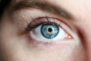
There are other atypical manifestations of cat-scratch disease that involve the eyes. Ophthalmic complications of cat-scratch disease include neuroretinitis, papillitis, optic neuritis, and Parinaud Oculoglandular syndrome. Khalf, N and Lin, D explain that while conjunctival infection may last only weeks, ‘the lymphadenopathy may persist for months.’ [10]
Left periocular pain is a chronic part of my condition. I have pain and swelling all around my eft eye, which is particularly severe around my temporal artery. There is definitely no conjunctival infection now, but I do wonder if this stubborn swelling and pain could be chronic lymphadenopathy.
Calcifications.
John Ghidoni, who wrote a research paper on the role of Bartonella henselae in the nucleation of calcification, confirms that Bartonella henselae does cause calcification in soft tissue. ‘Bacterial membranes contain calcium apatite crystals. Antigenic material was present in bacteria and calcified nodules. This case illustrates calcified probiotics becoming incorporated into scar tissue.’ [11]
The soft tissue that a surgeon removed in 2018 during my parotid gland surgery had calcifications within it. Unfortunately, they did not culture the tissue to try and ascertain what bacteria had caused the hardened tissue. It was found nonetheless. I am certain that there is still calcified soft tissue on the left side of my face and around my left eye, where I experience pain. It feels rock solid to touch. I can also see in the radiology images large areas of unexplained white on the left side of my face. I have been unable to get answers about whether this is calcified tissue.
My Radiology images.

Radiology image from CT neck scan, 2020. Red arrow points to the white area on my affected side.
In my radiology images from an MRI neck scan (2022) and a CT neck scan (2020), there are areas of white on the left side of my face and around my left eye. The dense white areas that are visible on the left side of my face are not visible on my normal side. I have been trying to ascertain if these white areas are calcified tissue. If this is calcified tissue, is Bartonella henselae the cause?
Treatment.

Medical professionals recommend antibiotic treatment for Bartonella infection in order to reduce the duration of the disease, particularly in cases of severe or chronic pain. Antibiotic treatments that doctors use to treat Bartonella infection include rifampicin, ciprofloxacin, trimethoprim-sulfamethoxazole, and gentamicin.
I have tried oral rifampicin and two of the fluoroquinolones: Levaquin and Ciprofloxacin. These were effective in softening my pain. I have not tried gentamicin or trimethoprim-sulfamethoxazole. I have asked numerous times for doctors to administer intravenous (IV) gentamicin or vancomycin, but they have repeatedly said no.
Reflection.

The suspected aetiology of my illness is infection and inflammation. Bartonella henselae seems like a strong possibility as a diagnosis. Bartonella henselae is transmitted to humans by fleas and cat scratches. A fleabite to my left eye triggered my illness. Bartonella infection/cat scratch disease could account for the calcified tissue that doctors have found in my soft tissue, for my chronic lymphadenopathy, and for the involvement of my swollen parotid gland.
Left peri-ocular pain and severe pain in my parotid gland are two chronic and painful aspects of my condition. Could both my unexplained parotid gland and ophthalmic involvement could be atypical manifestations of CSD? Cat scratch disease could explain why my illness has gone undiagnosed for so long, as Bartonella hensela can go undetected for decades.
There have been clinical descriptions of cat scratch disease for over 50 years, but the bacteriologic agent was not identified for decades. [12]
Conclusion.


The persistent clinical evidence over the years relating to my condition has been raised neutrophils, WBC, and MCHC. Many research papers on Bartonella infection state that lymph node biopsy should be considered if diagnosis is unclear. I don’t understand why clinicians have never done a serology or a lab culture of my lymph node tissue. They could have cultured my lymph nodes in 2018, when the oral maxillofacial surgeon excised my parotid tissue, but they didn’t. It would be useful if medics could still perform this investigation.
Lymph node biopsy is not indicated routinely but should be considered if the diagnosis is unclear.[13]
I think the person who reached out to me on social media may be correct. I think that I may well have a Bartonella infection. Research shows that Bartonella infection causes tissue to calcify and harden. Medics have found calcified deposits in my tissue. I worry that further calcium deposits could be causing my head pain. If abnormal tissue is the cause, I am concerned that it will be too late for doctors to treat this.
I am actively seeking an infectious disease specialist who has an interest in chronic infections in the head and neck and who could look into whether Bartonella henselae is a possible cause of my facial pain. I would welcome any contact from an ID clinician from anywhere in the world.
Cat-scratch should be considered in the differential diagnosis of any acute, subacute, or chronic lymphadenopathy. [14]

Foot Notes
[1] Koirala J, MD, Sunadareshan V, MD, and Basnet S, MD: Bartonella Infection, BMJ Best Practice, June 2023, www.bmj.com
[2] This information is from the same source as footnote 1.
[3] Rombaux P et al. Cervical lymphadenitis and cat scratch disease (CSF): an overlooked disease? PubMed.
[4] Ridder G J et al. Cat-scratch disease: Otolaryngologic manifestations and management, PubMed, March, 2005.
[5] Dreher A and Grevers G: Cat-scratch disease. An overview for the ENT physician, PubMed, July 1996.
[6] Ridder G J et al. Cat-scratch disease: Otolaryngologic manifestations and management, PubMed, March 2005.
[7] Ridder G J et al. Parotid involvement in cat scratch disease: a differential diagnosis with increased significance, National Library of Medicine (NIH), August 2000.
[8] This quotation is from the same source as footnote 7.
[9] Rombaux P et al. Cervical lymphadenitis and cat scratch disease (CSD): an overlooked disease? 2005.
[10] Khalf N and Lin D: Diagnosis and interpretation of testing for cat scratch disease, Copyright © 2021 Baylor University Medical Centre, Taylor and Francis Group.
[11] Ghidoni, J.: Role of Bartonella henselae endocarditis in the nucleation of aortic valvular calcification, February 2004, National Library of Medicine (NIH).
[12] Baranowski K and Huang B: Cat Scratch Disease, StatPearls; January 2024.
[13] Khalfe N and Lin D: Diagnosis and interpretation of testing for cat scratch disease, Copyright © 2021 Baylor University Medical Centre.
[14] Ridder G J et al. Cat scratch disease: Otolaryngologic manifestations and management, March, 2005, National Library of Medicine (NIH).
Useful Videos
Photo Credits: Edward Jenner, Peyma Farmani, CDC, National cancer Institute and 42 North on unsplash. Shvets Production on Pexels. Anja from Pixabay.
I Really appreciate your blog. AS I’M TREATING b
Bartonella, Babesia and Lyme. Have you cured your infection yet? How are You Doing, Friend?
Hi Anita, Thank you for your comment and feedback. No, I have sadly had no cure for my infection yet. Best wishes, Felicia